Home
/ Arteries In Neck Diagram / Vascular Anatomy Of The Neck Ent Clinic Sydney / In severe cases, a stroke can be fatal.
Arteries In Neck Diagram / Vascular Anatomy Of The Neck Ent Clinic Sydney / In severe cases, a stroke can be fatal.
Arteries In Neck Diagram / Vascular Anatomy Of The Neck Ent Clinic Sydney / In severe cases, a stroke can be fatal.. Despite being a relatively small region, it contains a range of important anatomical features. Figure schematic owchart from the arteries in the neck and the external carotid carries blood to structures outside the skull primarily the face. The stapedial artery connects the internal carotid and external carotid arteries. Smartdraw includes 1000s of professional healthcare and anatomy chart templates that you can modify and make your own. In this image, you will find external carotid artery, internal carotid artery, vertebral artery, aorta and arch, pulmonary artery, cardiac artery, thoracic aorta, celiac trunk, superior mesenteric artery, renal artery, gonadal artery, inferior mesenteric artery, common iliac artery, external iliac artery.
Figure schematic owchart from the arteries in the neck and the external carotid carries blood to structures outside the skull primarily the face. There are several head and neck arteries: The main artery in the neck is the common carotid artery, which divides at the upper border of the thyroid cartilage of the larynx (c4). The left and right common carotid arteries ascend up the neck, lateral to the trachea and the oesophagus. Contain the common carotid artery, internal.
Vascular Endovascular Surgery Carotid Endarterectomy from vascularsurgery.ucsf.edu Human body artery diagram in detail. Neck in land vertebrates the portion of the body joining the head to the shoulders and chest. In this image, you will find external carotid artery, internal carotid artery, vertebral artery, aorta and arch, pulmonary artery, cardiac artery, thoracic aorta, celiac trunk, superior mesenteric artery, renal artery, gonadal artery, inferior mesenteric artery, common iliac artery, external iliac artery. The left common carotid artery branches directly off the aortic arch and extends into the neck. Ninja nerds!join us in this video where we discuss the blood circulation of the head and neck using a flow chart. At the level of the superior margin of the thyroid cartilage (c4), the carotid arteries split into the external and internal carotid arteries. « back show on map ». Contain the common carotid artery, internal.
The content of the neck is grouped into 4 neck spaces, called the compartments.
A little above its termination is a second dilatation the. It can occur in the carotid artery of the neck as well as other arteries. The left and right carotid arteries each divide in the neck to form the left and right internal carotid as well as the left and right external carotid arteries. Start studying arteries of the neck and head. At the level of the superior margin of the thyroid cartilage (c4), the carotid arteries split into the external and internal carotid arteries. In this image, you will find external carotid artery, internal carotid artery, vertebral artery, aorta and arch, pulmonary artery, cardiac artery, thoracic aorta, celiac trunk, superior mesenteric artery, renal artery, gonadal artery, inferior mesenteric artery, common iliac artery, external iliac artery. Neck in land vertebrates the portion of the body joining the head to the shoulders and chest. Contain the common carotid artery, internal. The left common carotid comes directly off the aortic arch, while the right common carotid comes from the brachiocephalic. The procedure is conducted on medium to large arteries, like the carotid artery (neck), or femoral artery (leg). For more details go to edit properties. The right common carotid artery has a different initial course. It is a branch of the brachiocephalic trunk.
The stapedial artery connects the internal carotid and external carotid arteries. « back show on map ». The external jugular vein (v. The left common carotid comes directly off the aortic arch, while the right common carotid comes from the brachiocephalic. Contains cervical vertebrae and postural muscles.
Arteries Of The Neck And Head Human Anatomy Organs from www.medicalook.com Left and right common carotid. Contains cervical vertebrae and postural muscles. The left common carotid artery branches directly off the aortic arch and extends into the neck. Neck in land vertebrates the portion of the body joining the head to the shoulders and chest. The vessels transport a fluid. The external jugular vein (v. Related posts of arteries in the neck picture veins and arteries of the neck. Contains glands ( thyroid, parathyroid, and thymus ), the larynx, pharynx and trachea.
Human body artery diagram in detail.
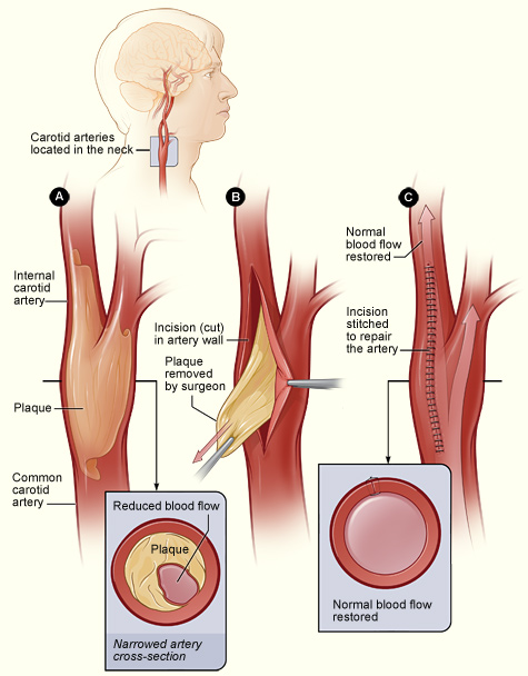
External carotid artery (supplies blood to the face and scalp) and and internal carotid artery. Neck in land vertebrates the portion of the body joining the head to the shoulders and chest. The carotid arteries extend out from the aorta artery, which transports blood out of the heart and is the body's largest artery. The left common carotid artery branches directly off the aortic arch and extends into the neck. A little above its termination is a second dilatation the. Head and neck arteries (diagram) blood supply for the head and neck comes from the branches of the aortic arch: Arteries of the head and neck the common carotid arteries supply blood to the head and neck. Smartdraw includes 1000s of professional healthcare and anatomy chart templates that you can modify and make your own. The procedure is conducted on medium to large arteries, like the carotid artery (neck), or femoral artery (leg). An endarterectomy is done under local or general anesthetic. Carotid artery disease causes about 10 to 20 percent of strokes. The brachiocephalic trunk gives rise to the right common carotid and right subclavian arteries. Instant anatomy is a specialised web site for you to learn all about human anatomy of the body with diagrams, podcasts and revision questions
At the level of the superior margin of the thyroid cartilage (c4), the carotid arteries split into the external and internal carotid arteries. The stapedial artery connects the internal carotid and external carotid arteries. The left and right common carotid arteries ascend up the neck, lateral to the trachea and the oesophagus. The right common carotid artery has a different initial course. Brachiocephalic trunk, left common carotid artery and left subclavian artery.
Vertebral And Internal Carotid Arteries Reprinted With Permission By Download Scientific Diagram from www.researchgate.net The cardiovascular system of the head and neck includes the vital arteries that provide oxygenated blood to the brain and organs of the head including the mouth and eyes. External carotid artery (supplies blood to the face and scalp) and and internal carotid artery. Related posts of arteries in the neck picture veins and arteries of the neck. The carotid arteries extend out from the aorta artery, which transports blood out of the heart and is the body's largest artery. The brachiocephalic trunk gives rise to the right common carotid and right subclavian arteries. The left common carotid artery branches directly off the aortic arch and extends into the neck. The left and right carotid arteries each divide in the neck to form the left and right internal carotid as well as the left and right external carotid arteries. A little above its termination is a second dilatation the.
It is a branch of the brachiocephalic trunk.
Each artery is a muscular tube lined by smooth tissue and has three layers: The left common carotid artery branches directly off the aortic arch and extends into the neck. Anterior view of internal anatomy of neck region hyoid bone trachea and thyroid. Ninja nerds!join us in this video where we discuss the blood circulation of the head and neck using a flow chart. The right common carotid artery has a different initial course. In severe cases, a stroke can be fatal. The trigeminal artery and hypoglossal arteries connect the blood vessels of the front and back of the head and neck. The artery is accessed through an incision in the neck or leg and the atheromatous plaque is physically removed usually as one piece with a spatula. The left common carotid comes directly off the aortic arch, while the right common carotid comes from the brachiocephalic. Veins and arteries of the neck 9 photos of the veins and arteries of the neck activate javascript arteries in the neck diagram, common carotid artery branches, external carotid artery function, how many carotid arteries, left common carotid artery function, the left common carotid artery supplies blood to the. Instant anatomy is a specialised web site for you to learn all about human anatomy of the body with diagrams, podcasts and revision questions The carotid arteries extend out from the aorta artery, which transports blood out of the heart and is the body's largest artery. A little above its termination is a second dilatation, the inferior bulb.
Contain the common carotid artery, internal arteries in neck. Learn vocabulary, terms, and more with flashcards, games, and other study tools.

